Current and Future Projects
The 353 LLC develops software applications and digital products across multiple industries.
Our Product Categories
Additional product information will be published as products become available.
EchoScholar
The only cardiac echo board prep app that asks you to identify structures directly on real echocardiographic images — the same way you think at the scanner.
Built the way echocardiographers actually think
Tap the structure directly on a real echo image. Identify anatomic landmarks, measurement points, and pathologic findings — spatially, not by process of elimination. No other cardiac echo board prep app offers this.
Cardiac tamponade, systolic anterior motion, sinus of Valsalva rupture, pulmonary embolism, aortic dissection on TEE, and more. Every cine and image is paired with a question, full explanation, and peer-reviewed citation.
All 1,000 questions include detailed explanations referenced to ASE guidelines, ACC/AHA documents, and peer-reviewed journals. Study from the source — not a summary.
1,000 Questions Across 5 Exam Domains
Mapped to ARDMS RDCS, CCI RCS, and ASE credentials — every domain tested on cardiac echo board examinations.
Every mode you need to be ready on exam day
Focused review by domain or across all 1,000 questions
Full-length timed simulation with instant scoring
Active recall for key facts and answer recognition
Reinforces weak areas using spaced repetition logic
Save any question to revisit later
Real questions from EchoScholar
This is exactly what you'll encounter in the app — real clinical media, real question formats, and full cited explanations.
Review these paired parasternal short axis cines at the level of the aortic valve. The 2D view shows disruption at the base of the aorta, and the color Doppler view reveals an abnormal high-velocity jet directed into an adjacent cardiac chamber. What is the diagnosis?
Show Explanation
A ruptured sinus of Valsalva (SOV) aneurysm produces a continuous shunt from the high-pressure aorta into an adjacent low-pressure chamber. The right coronary sinus most commonly ruptures into the right ventricle; the non-coronary sinus most commonly ruptures into the right atrium. Color Doppler demonstrates a characteristic continuous, high-velocity jet at the aortic root level — not at the interventricular septum as in a VSD. The clinical presentation is often acute onset of chest pain and a continuous 'machinery' murmur. The finding is distinct from aortic regurgitation, which produces a diastolic jet directed into the LVOT.
ASE Guidelines for Performing a Comprehensive Adult TTE: Mitchell C et al., J Am Soc Echocardiogr. 2019;32(1):1–64.
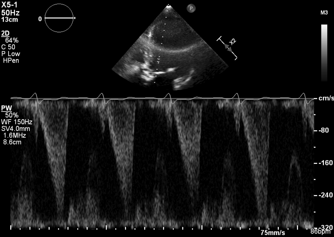
This CW Doppler tracing from the apical 5-chamber view demonstrates a late-peaking, concave, 'dagger-shaped' systolic signal in the LVOT. How does this waveform morphology differ from valvular aortic stenosis, and what diagnosis does it indicate?
Show Explanation
The CW Doppler waveform morphology is a key differentiator between fixed and dynamic LVOT obstruction. In HOCM, the obstruction is dynamic — it increases progressively during systole as the ventricle empties and the LVOT narrows due to SAM. This produces a characteristic late-peaking, concave, dagger-shaped waveform. In valvular aortic stenosis, the obstruction is fixed, producing a rounded, symmetric waveform that peaks in early-to-mid systole. The dagger shape is pathognomonic for dynamic obstruction. Additionally, HOCM gradients increase with provocative maneuvers that decrease preload (Valsalva, standing) and decrease with maneuvers that increase preload (squatting, passive leg raise).
AHA/ACC Hypertrophic Cardiomyopathy Guidelines: Ommen SR et al., J Am Coll Cardiol. 2020;76(25):e159–e240.
Simple, flexible subscriptions
Pricing designed with students in mind. Serious exam prep at a price that fits your life.
Available on iOS. Cancel anytime through your App Store account settings.
EchoScholar is an independent educational product developed by The 353 LLC. It is not affiliated with, endorsed by, or approved by ARDMS, CCI, ASE, or any credentialing organization. Content is intended for examination preparation only and does not constitute clinical guidance or medical advice.
"Product details and availability will be announced as development milestones are reached."
For inquiries about partnerships, licensing, or early access opportunities, contact us.